Ultrasound for Pets
Ultrasonography at Wigram Vets
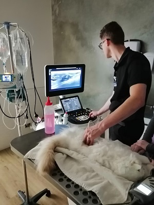
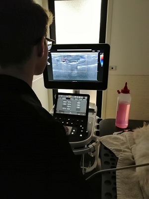
Ultrasonography is a non-invasive method of assessing the appearance and health of internal organs. The ultrasound converts reflected sound waves into real-time images, allowing for a detailed view of the inner workings of the body.
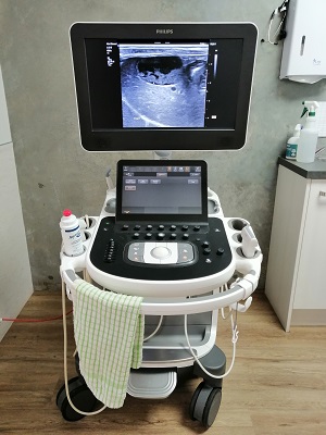
Unlike an X-ray which provides a static image, ultrasound captures a moving picture of the internal structure of an organ. This makes it a preferred option in many cases. Our clinic is equipped with a state-of-the-art ultrasound machine, acquired in 2024, that aids in accurate diagnosis and treatment guidance for a variety of conditions
Ultrasound can also be used to obtain needle aspirates from any abnormal organs found during the exam. This is a non-painful procedure which can help give us answers without the need for surgery.

Ultrasound is most commonly used for examination of abdominal organs. Some examples where an ultrasound can provide valuable insights include:
- Examination of the liver and gall-bladder in patients with elevated liver and bilirubin levels on blood tests
- Examination of the spleen for bleeding tumors
- Checking the size and appearance of the kidneys
- Diagnosing pancreatitis
- Checking for enlarged abdominal lymph nodes
- Assessing adrenal glands
- Bladder masses and stones
- Intestinal conditions
- Checking any abnormal fluid accumulations within the abdomen
- Diagnosing pregnancy and checking puppies if there are any abnormalities during birthing
Most patients will require a light sedation so they are relaxed and stay still for the procedure and can go home the same day.


